

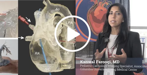
3D modeling, 3D printing and virtual reality (VR) are becoming more and more useful in the visualization of complex congenital and structural heart anatomies, offering clinicians ways to move beyond traditional 2D imaging.
Cardiovascular Business spoke with Kanwal Farooqi, MD, a pediatric cardiology imaging specialist and associate professor of pediatrics at Columbia University Irving Medical Center, about how her 3D lab helps add this extra dimension to patient care. She spoke about this very topic at SCCT2026, the annual meeting of the Society of Cardiovascular Computed Tomography (SCCT).
Farooqi described how her team uses 3D models to improve the visualization of challenging cardiac anatomies and support clinical decision-making, education, procedural planning and peri-procedural guidance.
She stressed that not every patient requires a physical model. In many cases, a digital 3D reconstruction provides the necessary visualization, allowing clinicians to crop the model in different planes and manipulate the anatomy on a computer screen. But for selected complex cases, the digital model can be converted into a physical 3D-printed representation. Farooqi said these models can provide a more intuitive way for clinicians, trainees, surgeons and families to understand anatomy that can be difficult to interpret on conventional 2D CT images.
